PUCRS researchers joined study to analyze lung images to aid diagnosis of Covid-19
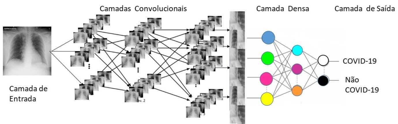
Image 1: Artificial convolutional neural network ranking lung radiography images
A multidisciplinary group from the University, in collaboration with researchers from the Universidade Federal de Uberlândia (UFU), is studying lung x-rays to aid in the diagnosis of Covid-19.
As the virus attacks the lungs and causes pneumonia, lung images such as chest radiography and computed tomography can assist doctors in the diagnosis and even in the prognosis of the disease.
According to Professor Dr. Ana Maria Marques da Silva, coordinator of PUCRS’ Laboratory of Computing in Medical Images, the objective of the investigation, which has already been approved by the University’s Ethics Committee, is to spot the differences between individuals who tested positive for Covid-19 and other infectious and inflammatory lung diseases. The findings will help researchers study the effects of Covid-19 on the lungs.
“In more advanced stages, images can indicate the extent or progress of the disease in the lungs at different times of treatment. However, in the early stages of the disease, these changes can be quite subtle, as it the speed of progress can vary or may not have any serious effects on the lungs. Thus, little is known about how imaging tests can predict the severity of the disease (prognosis). Our research aims to investigate exactly how computer-based radiological images, known as radiomic signature, can be associated with the prognosis and severity of the disease ”, she explains.
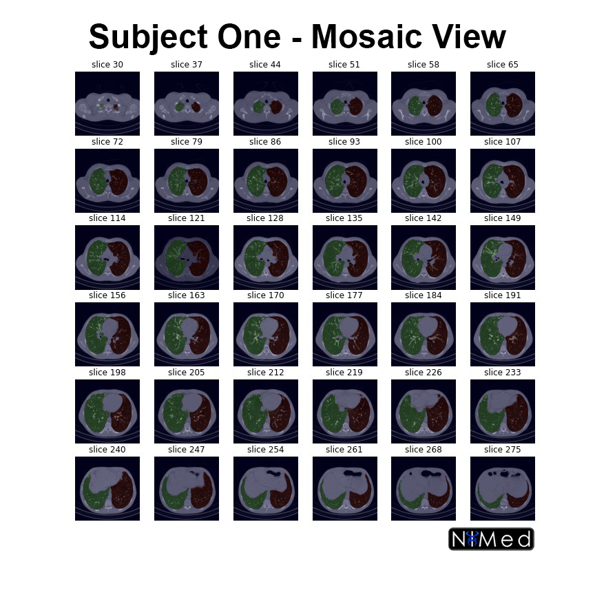
Image 2: Lung segmentation on chest x-rays. The tool separates the lungs and identifies the region of the heart. This ensures that the analysis will look at no other region in the body
In the beginning of the winter, people are more susceptible to lung diseases, which leads a greater number of imaging tests in medical facilities to check for lung problems. In view of that, the researcher envisions the use of more images for research. This will also increase the reliability of the results.
She anticipates that when the first phase of studies with lung x-rays is completed, the group will be able to provide a computational tool that should help doctors identify lung images with Covid-19. In the second phase, using CT-scans, the analysis can be improved and extended to indicate stages of the disease.
Dr. da Silva points out that no tool will completely replace tests to identify Covid-19, but as they emerge, they will be able to help rank patients in view of their likelihood to have their condition aggravated in an effort to provide better target treatments.
At the Universidade Federal de Uberlândia (UFU), an artificial neural network has been developed (Figure 1) to tell radiographic images of healthy individuals from those of individuals with Covid-19. According to the researchers, the neural network used hundreds of lung X-ray images that are being made available in public databases created with radiographs from all over the world.
At PUCRS, x-rays are being collected from patients diagnosed with Covid-19 to assess the artificial neural network and increase its reliability. An automatic lung segmentation tool has been implemented on x-rays (Figure 2) and computed tomography (Figure 3). The university is working on the validation of these segmentations.
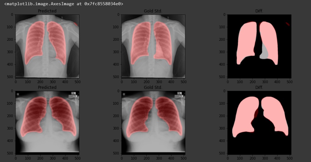
Image 3: Lung segmentation results on chest CT scans. The tool separates the left and right lungs in red and green for analysis
In addition, the team of researchers, including physicists, engineers, computer scientists and physicians, is also collecting as many x-rays and CT scans as possible from diagnosed individuals who are undergoing treatment or have had different disease progressions. According to the project leader at PUCRS, knowing how patients evolved over the course of the disease will allow them to investigate which radiomic characteristics of the images can give an indication of the evolution of the disease.
At PUCRS, the project is led by Professor Dr. Ana Maria Marques da Silva, coordinator of the Laboratory of Computing in Medical Images, with the participation of professors Dr. Bruno Hochhegger and Dr. Rodrigo Coelho Barros, as well as undergraduate and graduate students. At UFU, the research is led by Professor Dr. Ana Cláudia Patrocínio, from the Biomedical Engineering Laboratory, with a team of undergraduate and graduate students.


